Search by last, first or email
Research Area
Departments
UCI School of Medicine
UCI School of Biological Sciences
 Abbott, Geoff W., PhD
Mailing Address: Research in Dr. Abbott’s lab is focused on elucidating the molecular basis for ion channel and transporter physiology and pathophysiology. One of the main directions of the lab is understanding the molecular basis for our recent discovery that some neurotransmitters can directly activate voltage-gated potassium channels. Another current emphasis is investigating the molecular basis of action of botanical folk medicines, arising from our recent findings that KCNQ potassium channels are an important target for plant metabolites. The Abbott lab also continues to uncover the roles of the KCNQ and KCNE families of ion channel subunits, using a combination of techniques including mouse and human genetics, electrophysiology, pharmacology, and state-of-the-art imaging modalities. A further area of focus is studying the physiological roles of recently discovered macromolecular complexes comprising KCNQ family Kv alpha subunits and sodium-coupled solute transporters. Abnormal functioning of ion channels causes many different disorders, including cardiac arrhythmia, diabetes, epilepsy, myotonia and periodic paralysis. Dr. Abbott’s lab uses a multidisciplinary approach aimed at understanding the molecular bases for these “channelopathies” and also how these proteins function normally to enable the complex signaling and cellular processes required by living organisms.
...view more
Research Areas: Drug Addiction, Neuropharmacology & Neurochemistry, Neurodegeneration & Neuropsychiatric Disorders |
 Acharya, Munjal, PhD
Mailing Address: Our laboratory focuses on neurobiological mechanisms and regenerative medicine approaches to treat cancer and cancer therapy-related cognitive impairments (CRCI). We use molecular, cellular, genetic, and behavioral techniques to focus on the following pre-clinical research emphases: 1) Glial complement cascade signaling mechanism in cranial radiation therapy and glioblastoma-induced neuroinflammation and cognitive dysfunction (supported by NIH). 2) Astrocyte-dependent mechanism of radiation-induced cognitive impairments and disruption of circadian rhythm (supported by American Cancer Society) 3) Human neural stem cell-based regenerative approaches to treat radiation- and chemotherapy-induced cognitive decline and synaptic damage (supported by CIRM).
...view more
Research Areas: Learning & Memory, Neurodegeneration & Neuropsychiatric Disorders, Neuroimmunology, Neuroinflammation & Glia |
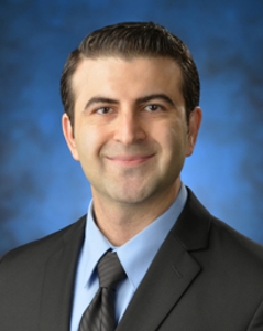 Akbari, Yama, M.D., Ph.D.
Mailing Address: Coma, Consciousness, Cardiac Arrest; Optical Imaging of cerebral blood flow and brain metabolism; optimizing CPR; stroke; traumatic brain injury; neural monitoring devices; translational research; clinical trials
...view more
Research Areas: Human Neuroscience |
 Anderson, Aileen J., PhD
Mailing Address: My laboratory focuses on a combination of discovery biology and identifying translational neuroscience strategies for spinal cord injury and central nervous system disease. Pre-clinical and translational work from my lab directly supported an IND filing for an FDA approved phase I trial of human neural stem cells for Pelizaeus-Merzbacher disease (PMD), and a phase I/II clinical trial for human neural stem cells in thoracic spinal cord injury (SCI). These translational milestones reflect our work on human-derived stem cell populations and spinal cord injury mechanisms. In current studies, we are investigating the intrinsic and extrinsic factors defining the migration and differentiation potential of these cells both in vitro and after in vivo transplantation. In parallel, we are seeking to understand non-traditional roles for the innate inflammatory system in the pathophysiology of spinal cord injury and in the mechanisms controlling stem cell fate and migration. A third line of research focuses on implanted biomaterial scaffolds and engineered nanoparticles, which we have shown can provide an environment supporting robust axonal regeneration.
...view more
Research Areas: Neurodevelopment, Stem Cells & Plasticity/Regeneration, Neuroimmunology, Neuroinflammation & Glia |
 Baram, Tallie Z., MD,PhD
Mailing Address: Our past experiences influence who we are. In the Baram lab we focus on early-life events, and especially early-life adversity (ELA) or stress; we investigate the consequences of early-life experiences and probe how they influence brain molecules, cells and circuits and how these changes may result in cognitive and emotional problems. Using cutting edge methods, we find new regions (e.g., the paraventricular thalamus) where early-life experiences get encoded, and identify new brain projections, epigenetics mechanisms and circuit plasticity through which transient ELA impacts brain function long-term in a sex-dependent manner. We perform our studies in experimental animals, and then translate them back to human children and adults.
...view more
Research Areas: Genetics & Epigenetics, Human Neuroscience, Learning & Memory, Neurodegeneration & Neuropsychiatric Disorders, Neurodevelopment, Stem Cells & Plasticity/Regeneration, Systems/Circuit Neuroscience & Synaptic Transmission |
 Beier, Kevin, PhD
Mailing Address: My research is focused on identifying how experience modulates activity dynamics in neural circuits, both acutely and chronically. I aim to develop new methods and use these in combination with modern techniques such as optogenetics and in-vivo calcium imaging to understand the sources of adaptive and maladaptive plasticity that drive normal and pathophysiological behaviors. These include reward/aversion processing in normal appetitive behaviors, drug addiction, neurodegeneration, and neurodevelopmental disorders such as autism. I will also employ cutting-edge viral-genetic methods to map brain state-dependent activity in connected neural ensembles, and use this information to infer how our brain state drives us to perform particular behaviors. I also aim to take novel approaches to interrogating the sources of individual behavioral variation in neuropsychiatric conditions such as addiction, anxiety, and depression.
...view more
Research Areas: Drug Addiction, Neuropharmacology & Neurochemistry, Genetics & Epigenetics, Learning & Memory, Neurodegeneration & Neuropsychiatric Disorders, Systems/Circuit Neuroscience & Synaptic Transmission |
 Borrelli, Emiliana, PhD
Mailing Address: Our research focuses on the dopaminergic system and in particular on the signaling mediated by one of the dopamine receptor, the D2 receptor (D2R)
...view more
Research Areas: Drug Addiction, Neuropharmacology & Neurochemistry, Genetics & Epigenetics, Neurodegeneration & Neuropsychiatric Disorders, Systems/Circuit Neuroscience & Synaptic Transmission |
 Cahill, Larry, PhD
Mailing Address: Sex influences on brain function
...view more
Research Areas: Drug Addiction, Neuropharmacology & Neurochemistry, Genetics & Epigenetics, Human Neuroscience, Learning & Memory, Neurodegeneration & Neuropsychiatric Disorders, Neurodevelopment, Stem Cells & Plasticity/Regeneration, Neuroimmunology, Neuroinflammation & Glia, Systems/Circuit Neuroscience & Synaptic Transmission |
 Calof, Anne L., PhD
Mailing Address: Systems Biology of Stem Cells and Human Developmental Disorders–The Calof lab takes a systems biology approach to address questions in development, regeneration, and tissue homeostasis. We also work on vertebrate animal model systems to understand the etiology of human genetic diseases that affect development and function, especially of the cardiovascular/pulmonary system and nervous system.
...view more
Research Areas: Genetics & Epigenetics, Neurodevelopment, Stem Cells & Plasticity/Regeneration |
 |
 Chen. Lulu Y., PhD
Mailing Address: We aim to identify the key molecules and their signaling cascades involved in synapse formation and functions in learning and memory and their deficits in various disease conditions. We are particularly interested in the activity-dependent signaling pathways underlying synaptic structural changes. We study different synapse types in the brain, how these synapses form, function, and been regulated. We take advantage of genetic and molecular approaches which enable accurately target the specific types of neurons/synapses located at specific brain regions (spatial control) at specific development window (temporal control) to ask that how the synapses are developmentally regulated and how this regulatory mechanism fail in the disordered brains.
...view more
Research Areas: Learning & Memory, Neurodegeneration & Neuropsychiatric Disorders, Neurodevelopment, Stem Cells & Plasticity/Regeneration, Systems/Circuit Neuroscience & Synaptic Transmission |
 |
 Chrastil, Liz, PhD
Mailing Address: At the Spatial Neuroscience lab, we study human path integration, spatial memory, and large-scale navigation in complex environments. Our lab conducts experiments using both fully immersive virtual reality (at our CAVERN (Center for Ambulatory Virtual Environment Research in Navigation) Lab) and fMRI to understand how humans process self-motion information when navigating with and without landmarks. We look to examine how active and passive navigation affect learning a new environment applying these same techniques. In addition, we are interested in questions of how proprioceptive input, vestibular information, decision-making, and attention contribute to learning different types of spatial knowledge. Our lab is broadly interested in individual differences in navigational abilities. Our research examines the relationship between performance and brain function, looking at both brain structure and fMRI activation across individuals.
...view more
Research Areas: Human Neuroscience, Learning & Memory |
 Cramer, Karina S., PhD
Mailing Address: Neural functions rely on highly ordered circuit assembly during development. In the central auditory system, these connections must be very precise to allow for the fast and reliable processing needed in auditory perception. Our laboratory studies the development and plasticity of auditory brainstem circuits needed to localize sounds. We are interested in discovering the mechanisms that guide auditory axons to their correct synaptic targets and form specialized synapses. We are currently investigating this in three lines of research. Glial cells in auditory circuit formation. We are studying the contributions of glial cells, including astrocytes and microglia, to synapse formation and maturation in the auditory pathways. We have found that astrocytes are important for the formation of inhibitory synapses and for the formation of mature dendritic arbors. We are now exploring how microglia contribute to synapse formation and synaptic pruning in the auditory brainstem, and how these roles contribute to auditory function. These studies highlight the importance of cellular communication between neurons and glia during development. Caspase-3 in auditory brainstem development. While it is well-established that caspase-3 is an effector of cell death, studies have suggested additional roles in development. We found that active (cleaved) caspase-3 displays an intriguing expression pattern along the auditory pathway during development. It is initially seen in auditory nerve axons projecting centrally. Expression seems to shift to sequentially higher levels of auditory centers. Notably, transitions in expression occur at about the time synapses form between transition areas. When caspase-3 was inhibited during development, we observed significant axon targeting errors and defects in formation of auditory centers. Caspases are proteases that cleave other proteins. We are currently exploring which proteins are caspase-3 substrates during development and how they act to shape auditory circuits. Auditory brainstem dysfunction in neurodevelopmental disorders. Fragile X syndrome (FXS), a neurodevelopmental disorder associated with intellectual disability, is also frequently associated with hyperacusis, the perception that sounds of normal intensity are uncomfortably or painfully loud. We have explored how impaired auditory brainstem development might contribute to hyperacusis in FXS.
...view more
Research Areas: Neurodevelopment, Stem Cells & Plasticity/Regeneration, Neuroimmunology, Neuroinflammation & Glia, Systems/Circuit Neuroscience & Synaptic Transmission |
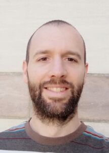 Diaz Alonso, Javier, PhD
Mailing Address: Throughout the brain, neurons communicate with each other through synapses, where neuronal specializations—presynaptic terminals, equipped with neurotransmitter release machineries, and postsynapses, containing neurotransmitter receptors—are connected by a narrow synaptic cleft. The correct positioning of elements in these three compartments allows for the flow of information between neurons. A series of phenomena referred to as synaptic plasticity can modulate synaptic strength, which endows us with the ability to learn from and adapt to a changing environment. In our lab we combine biochemistry, molecular neurobiology and electrophysiology to understand the molecular mechanisms that govern synapse development, organization and plasticity. Our goal is to decipher how signaling elements of the widespread glutamatergic and endocannabinoid systems are organized at synapses, how this organization is dynamically regulated to enable synaptic plasticity and how its disruption can lead to pathological states.
...view more
Research Areas: Learning & Memory, Neurodegeneration & Neuropsychiatric Disorders, Neurodevelopment, Stem Cells & Plasticity/Regeneration |
 Ewell, Laura, PhD.
Mailing Address: Cellular mechanisms of memory and epilepsy In the Ewell Lab, we strive to uncover the network mechanisms of learning and memory in health and disease. We focus on temporal lobe epilepsy – a disease characterized by spontaneous seizures and profound memory impairments. We employ large scale in vivo recording techniques to record from hippocampal networks duringmemory tasks. We have established that epileptic mice have deficits in spatial working memory, and that they have multiple alterations in the neural codes that support this type of memory. We are currently developing novel automated high throughput behavioral tasks to probe the impact that such impaired dynamics have on encoding, consolidation, and recall of spatial memories.
...view more
Research Areas: Learning & Memory, Systems/Circuit Neuroscience & Synaptic Transmission |
 Flanagan, Lisa A., PhD
Mailing Address: Dr. Lisa Flanagan is a member of the Department of Neurology at the University of California, Irvine, with joint appointments in Biomedical Engineering and Anatomy and Neurobiology. Dr. Flanagan’s research program combines the fields of neural cell biology and bioengineering to address issues critical to the successful transplantation of neural stem cells (NSCs) for the repair of injured or diseased central nervous system (CNS) tissue. As such, her lab is investigating factors that affect the differentiation of NSCs into the three CNS lineages (neurons, astrocytes or oligodendrocytes) and exploring means to direct fate decisions of transplanted or endogenous cells for optimal repair and regeneration. Dr. Flanagan’s lab has taken two complementary approaches to these problems. In one, they are developing novel non-invasive methods to identify the fate potential of undifferentiated stem cells with the goal of isolating cells biased to a particular fate. In the other, they investigate the role of extracellular matrix cues in regulating the behavior of NSCs in order to generate instructive three-dimensional matrices for NSC transplantation. Their goal is to achieve greater control over the differentiation of transplanted stem cells to unravel the contributions of each cell type to effective repair and potentially maximize functional recovery. These studies have clinical applications for the treatment of stroke, Alzheimer’s disease, and spinal cord injury.
...view more
Research Areas: Neurodevelopment, Stem Cells & Plasticity/Regeneration |
 Fortin, Norbert J., PhD
Mailing Address: The main goal of our lab is to understand the fundamental neural mechanisms underlying our ability to temporally organize our memories, as well as how this capacity is affected in cognitive disorders. Our primary approach is to train rats to perform complex sequence memory tasks (comparable to those used in humans) and then use high-density electrophysiological recordings techniques to examine how ensembles of neurons represent and compute the information needed to solve the task. Notably, unlike most rodent labs, we work in close collaboration with human fMRI labs to establish the cross-species correspondence of our research (at the behavioral and neural level). We also work in close collaboration with colleagues in Statistics to develop the analytical tools necessary to “decode” the information represented in the neural activity. At this time, we primarily focus on the fundamental function of the hippocampus and prefrontal cortex, as well as their functional interactions with associated structures, but are in the process of extending these approaches to study the neural mechanisms underlying memory impairments associated with aging, dementia, hypoxia, and radiotherapy.
...view more
Research Areas: Learning & Memory, Systems/Circuit Neuroscience & Synaptic Transmission |
 Fowler, Christie D., PhD
Mailing Address: The overall goal of my research is centered on elucidating the genetic, epigenetic and molecular mechanisms underlying motivated behaviors that characterize drug addiction. My research team conducts cutting-edge studies to define the pathways and mechanisms regulating the reinforcing and aversive processing of nicotine and cannabis. Our multi-dimensional approach employs innovative techniques, along with well-established molecular and behavioral analyses, to more clearly reveal the specific role of signaling molecules, neurotransmitters and neural circuits involved in drug-induced changes in neuronal function. Based on the increasing incidence of e-cigarette and cannabis use among adolescents, we also investigate the impact of developmental drug exposure (both single substance and co-exposure with cannabinoids and nicotine) on later drug taking, cognitive and affective behaviors in adulthood. Most recently, we have also developed a novel area of investigation to define the role of circulating extracellular vesicles (exosomes) on neural function across the substance use trajectory. Finally, through several collaborations with medicinal chemists in the US and Europe, we conduct pre-clinical testing of novel compounds to derive more efficacious therapeutic treatments to combat drug addiction.
...view more
Research Areas: Drug Addiction, Neuropharmacology & Neurochemistry, Genetics & Epigenetics, Learning & Memory, Neurodegeneration & Neuropsychiatric Disorders, Systems/Circuit Neuroscience & Synaptic Transmission |
 Frostig, Ron, PhD
Mailing Address: Research interest: Neocortical structure, function and plasticity at the large neuronal ensembles level: from basic to pre-clinical research. Neocortex, believed by Neuroscientists to be the hallmark of brain evolution, is still poorly understood and therefore constitutes a major challenge for Neuroscientists. This challenge calls for an innovative combination of new concepts about cortical function combined with state-of-the-art technologies.
...view more
Research Areas: Neurodevelopment, Stem Cells & Plasticity/Regeneration, Systems/Circuit Neuroscience & Synaptic Transmission |
 Gall, Christine M., PhD
Mailing Address: Research in the Gall laboratory is focused on the cellular mechanisms of synaptic plasticity underlying learning and memory. Recent work has addressed the hypothesis that a form of synaptic plasticity thought to underlie memory, referred to as long term potentiation (LTP), depends on both remodeling of the synaptic cytoskeleton and a precise balance in signaling from a number of synaptic modulatory receptors. In particular, we find that among relays in limbic circuitry encoding episodic memory, there are differences in mechanisms of plasticity with endocannabinoid signaling required at one point and sexually dimorphic involvement of estrogen at another. Parallel studies are evaluating impairments in synaptic plasticity in fragile X syndrome model mice and testing the efficacy of therapeutics targetting modulatory signaling (e.g., estrogen, endocannabinoid, oxytocin) to enhance cognitive function. Ongoing projects are testing the effects of early life cannabinoid exposure on synaptic and cognitive function in adulthood, evaluating mechanisms through which oxytocin improves cognitive function in fragile X model mice, and identifying the synaptic bases of sexual dimorphism in hippocampus dependent learning. Procedures include evaluating learning behavior, synaptic/slice electrophysiology and quantitative microscopy (to evaluate synaptic receptors and signaling).
...view more
Research Areas: Learning & Memory, Neurodevelopment, Stem Cells & Plasticity/Regeneration, Systems/Circuit Neuroscience & Synaptic Transmission |
 Gelinas, Jennifer, MD,PhD
Mailing Address: Epilepsy is more than just seizures.
...view more
Research Areas: Human Neuroscience, Learning & Memory, Neurodevelopment, Stem Cells & Plasticity/Regeneration, Systems/Circuit Neuroscience & Synaptic Transmission |
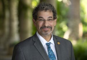 Goldstein, Steve A. N., MD,PhD
Mailing Address: Our research seeks to understand how ion channels operate in health and illness. Ion channels are membrane proteins in all cells that catalyze the selective passage of ions across membranes and, like enzymes, show exquisite specificity and tight regulation. As a class, ion channels orchestrate the electrical activity that allows operation of the heart, nervous system and skeletal muscles and are just as important in non-excitable cells like circulating T cells and sperm. Less sensational but equally important, ion channels mediate cellular fluid and electrolyte homeostasis. Remarkably, fundamental questions remain to be answered. How do ion channels open and close? What is their architecture? How do mutations produce cardiac arrhythmia, hypertension, seizures, or interfere with immune function? How do drugs act to produce beneficial outcomes (~20% of our current pharmacopeia targets ion channels) or to yield undesirable side effects? Our laboratory uses macroscopic and single molecule electrophysiology and spectroscopy, molecular genetics, high-throughput and structural methods to improve human health and wellness.
...view more
Research Areas: Drug Addiction, Neuropharmacology & Neurochemistry, Human Neuroscience, Neurodegeneration & Neuropsychiatric Disorders, Neuroimmunology, Neuroinflammation & Glia |
 Green, Kim, PhD
Mailing Address: Since starting my own lab in 2011, I have focused on microglia, the immune cell of the brain. We discovered that microglia in the adult brain are dependent upon signaling through the colony-stimulating factor 1 receptor (CSF1R) for their survival, and that we could take advantage of this dependency through the administration of specific CSF1R inhibitors leading to the rapid and sustained elimination of >95% of all microglia from the CNS. Through this method we are studying the roles that microglia play in normal brain function, as well as their effects on the brain during disease and injury. We also uncovered that once the brain has been depleted of microglia withdrawal of CSF1R inhibitors leads to the rapid repopulation of the entire CNS with new microglia within a few days. We are exploring the therapeutic implications eliminating microglia after injury, or in disease, and then replacing them with new cells. See our publications at:
...view more
Research Areas: Neurodegeneration & Neuropsychiatric Disorders, Neuroimmunology, Neuroinflammation & Glia |
 Grill, Joshua, PhD
Mailing Address: For any new Alzheimer’s disease drug to achieve FDA approval and widespread clinical use, it must be tested in human beings and demonstrated as both safe and efficacious. Studies to test the safety and efficacy of new treatments are called clinical trials. Clinical testing now represents the most costly and difficult phase of developing improved therapies. A major challenge of completing human clinical trials is the timely enrollment of participants who will enable adequate examination of therapeutic hypotheses. Alzheimer’s disease clinical trials now enroll people with Alzheimer’s dementia, people with mild cognitive impairment, and people with healthy memories but who are at increased risk to some day develop dementia. We are engaged in a variety of studies that aim to
...view more
Research Areas: Neurodegeneration & Neuropsychiatric Disorders |
 Head, Elizabeth, PhD
Mailing Address: I have been interested in learning and memory, with a focus on aging since I was a graduate student in Behavioral Neuroscience at the University of Toronto where I played a part in developing novel cognitive tasks to assess brain aging in an animal model of Alzheimer disease. These early studies in cognition formed the foundation for my current research program that subsequently developed at UCI, which is to characterize aging and the development of Alzheimer disease in people with Down syndrome. Through clinical longitudinal studies with a cohort of people with Down syndrome, my team links aging to changes in learning, memory and executive function to biomarker outcomes such as neuroimaging. Coupled with basic science research examining various molecular pathways engaged in the aging and Alzheimer disease process through autopsy studies, my lab seeks to identify targets for interventions that may improve cognition. I am delighted to have returned to UC Irvine to continue to build my research program.
...view more
Research Areas: Human Neuroscience, Learning & Memory, Neurodegeneration & Neuropsychiatric Disorders |
 Hunt, Robert, PhD
Mailing Address: My lab focuses on conditions of brain development and injury where a treatment does not yet exist. We are creating new models of complex neurological disorders, developing cellular and molecular tools that enable precise manipulation or repair of the brain and recording high-speed dynamics of specialized neurons derived from human stem cells. Our philosophy is to embrace a broad view of neuroscience as we integrate a constellation of molecular, cellular, developmental, electrophysiological and systems-level methods in our work. Ultimately, we hope to discover precisely how the nervous system is changed in neurodevelopmental disorders or by traumatic brain injury, and to use this information to create new therapies. Our research has been funded by NIH, NSF and private sources, including a K99/R00 Pathway to Independence Award from the National Institute of Neurological Disorders and Stroke. Here at UCI, the lab participates actively in the Interdepartmental Neuroscience Program, Epilepsy Research Center, Center for Autism Research and Translation, Stem Cell Research Center and the Center for the Neurobiology of Learning and Memory.
...view more
Research Areas: Genetics & Epigenetics, Learning & Memory, Neurodegeneration & Neuropsychiatric Disorders, Neurodevelopment, Stem Cells & Plasticity/Regeneration, Systems/Circuit Neuroscience & Synaptic Transmission |
 Igarashi, Kei M., PhD
Mailing Address: The Igarashi lab investigates brain circuits for memory in Health and Disease The scent of your parents’ signature gingerbread can trigger sweet childhood memories and will vividly remind you of past events. Impairments of memory function of the brain, on the other hand, directly lead to the disruption of our daily life. Alzheimer’s disease is currently affecting >50 million patients worldwide. Identifying brain circuit mechanisms underlying memory is not only a major goal for the basic neuroscience field, but also an ultimate goal for clinical neuroscience. The Kei Igarashi laboratory at UC Irvine investigates: To solve these problems, we are targeting the entorhinal-hippocampal circuits, a core brain circuit for memory. We use state-of-the-art systems neuroscience techniques including: These works are supported by grants from NIH/NIA, NIH/NIMH, Whitehall Foundation, Alzheimer’s Association, Brain Research Foundation, Donors Cure Foundation, and BrightFocus Foundation.
...view more
Research Areas: Learning & Memory, Neurodegeneration & Neuropsychiatric Disorders, Systems/Circuit Neuroscience & Synaptic Transmission |
 Jin, Rongsheng, PhD
Mailing Address: Our research group is dedicated to understand the molecular basis of human diseases using structural biology, which allows us to visualize how proteins function or malfunction at the atomic level. A major area of our research concerns the structure and function of botulinum neurotoxins (BoNTs), which are among the most poisonous substances known to humans. BoNTs therefore represent a major bioterrorist threat. Paradoxically, BoNT-containing medicines and cosmetics have been used with great success in clinic. Both the toxic and therapeutic functions of BoNTs indeed rely on a common mechanism to enter motoneurons, cleave SNAREs that mediate release of acetylcholine, and subsequently paralyze the affected muscles. We are trying to understand the molecular mechanisms underlying the BoNT-host interplay. These studies will help to improve clinical efficacy of BoNT-containing drugs, suggest novel therapeutic applications for BoNTs, and guide the development of effective anti-BoNT strategies. In parallel, we are striving to understand the structural mechanisms underlying synapse formation, neurotransmission, and synaptic plasticity, which may facilitate the design and improvement of therapeutic agents for the treatment of some psychological and neurological disorders.
...view more
Research Areas: Drug Addiction, Neuropharmacology & Neurochemistry, Systems/Circuit Neuroscience & Synaptic Transmission |
 Jusiak, Barbara, PhD
Mailing Address:
...view more
Research Areas: Uncategorized |
 Kefalov, Vladimir, PhD.
Mailing Address: Research Interests: Sensory Neurobiology, Photoreceptor physiology, Visual cycle and dark adaptation, Photoreceptor degeneration, Gene-independent therapy for visual disorders We are a sensory neurobiology lab interested in the function of mammalian rod and cone photoreceptors. Studies target basic biological questions such as cone and rod phototransduction, calcium and the mechanisms of light adaptation, and the visual cycle and dark adaptation. Additional studies focus on disease-oriented questions such as the mechanisms of retinal degeneration in mouse models of visual disorders, and the development of therapeutic tools for preventing photoreceptor death. Current projects investigate the signaling by chromophore-free opsin, chromophore supply to cones, the etiology of G90D and G90V rhodopsin mutations, and calcium regulation in rod bipolar cells.
...view more
Research Areas: Neurodegeneration & Neuropsychiatric Disorders, Systems/Circuit Neuroscience & Synaptic Transmission |
 La Spada, Al, PhD
Mailing Address: Dr. La Spada’s research is focused upon neurodegenerative disease and neurotherapeutics, and he is seeking the molecular events that underlie neurodegeneration and neuron cell death in spinal & bulbar muscular atrophy (SBMA), spinocerebellar ataxia type 7 (SCA7), Huntington’s Disease, ALS, and Parkinson’s disease. He and his team have uncovered evidence for transcription dysregulation, perturbed bioenergetics, and altered protein quality control as contributing factors to neuron dysfunction. His research is also focused upon cell-cell communication between neurons and non-neural cells, emphasizing the role of astrocytes and microglia in disease pathogenesis and progression in CNS disease, and the contribution of skeletal muscle in motor neuron diseases. By reproducing molecular pathology in mice and in neurons and other cell types derived from human patient stem cells, Dr. La Spada has begun to develop therapies to treat these disorders intended to boost the function of pathways of CNS homeostasis that decline with aging.
...view more
Research Areas: Genetics & Epigenetics, Neurodegeneration & Neuropsychiatric Disorders |
 LaFerla, Frank M., PhD
Mailing Address: The broad goal of my lab is to elucidate the pathophysiological mechanisms underlying neurodegenerative disorders such as Alzheimer’s disease and other dementias by developing and using novel animal models in parallel with studies from affected human subjects. The LaFerla laboratory has a long-standing interest in understanding how co-morbidities influence the onset and progression of Alzheimer’s disease. Sporadic AD patients typically display 2 to 8 different comorbid conditions, and it is possible that these concomitant medical conditions influence the progression of AD. Among the diversity of the different co-morbid conditions, the lab has focused on three: ischemia/stroke, stress and diabetes. Our findings have contributed significantly to understanding how stress and ischemia impact AD pathogenesis. Recently, we discovered that type 1 diabetes requires tau to induced synaptic and cognitive impairments. These observations have led us to investigate if the more common form of diabetes (type 2) also requires tau for the manifestation of the cognitive and synaptic impairments and to further explore the underlying molecular mechanisms by which synaptic integrity is compromised by type 1 and 2 diabetes. These studies have led to a better understanding of the molecular connections by which two highly prevalent human disorders are related, which could facilitate the development of novel interventions for AD patients with diabetes. Inflammation is a fundamental protective response, but if it becomes dysregulated, it can be a major co-factor in the pathogenesis of many chronic human diseases, including Alzheimer disease. My laboratory has made important discoveries as to how inflammatory processes trigger AD pathogenesis (including Ab and tau) and impair cognition. Currently, we are investigating the impact of Ab on IL-1b signaling, with an emphasis on the relevance of protein clearance for IL-1b synthesis and protein trafficking for IL-1R1 levels. The translational impact of this work is substantial and significant, as these studies have uncovered new therapeutic targets for the treatment of AD.
...view more
Research Areas: Neurodegeneration & Neuropsychiatric Disorders |
 Lane, Thomas E., PhD
Mailing Address: Work in my laboratory is divided into three main research areas: 1) define the functional contributions of chemokines and chemokine receptors in defense and disease following viral infection of the central nervous system (CNS), 2) evaluate the therapeutic potential of mouse/human neural precursor cells (NPCs) in clinical recovery and remyelination in a model of viral-induced demyelination and 3) study SARS-CoV2 pathogenesis in vitro and vivo. My laboratory has a long-standing interest in understanding events that initiate and maintain inflammation within the CNS in response to viral infection. To this end, we have set forth on a directed path to determine the functional significance of chemokines and chemokine receptors in both host defense as well as disease development following instillation of a positive-strand RNA virus (mouse hepatitis virus – MHV) into the CNS of susceptible mice. Indeed, we have shown that blocking chemokine function via both antibody neutralization and genetic silencing in virally-infected mice resulted in increased mortality accompanied by reduced immune cell infiltration into the CNS. Subsequently, we have demonstrated that unique chemokine/chemokine receptor signaling pathways are critical for interrelated events required for optimal host defense following viral infection including linking innate and adaptive immune responses, regulating antiviral effector functions e.g. cytokine secretion/cytolytic activity by effector T cells, and promoting the directional migration of antigen-sensitized lymphocytes into the CNS. We have also focused on how chemokine signaling influences the biology of oligodendroglia with regards to protection from inflammatory cytokine-induced apoptosis. Similar to the human demyelinating disease multiple sclerosis (MS), remyelination failure is also observed in MHV-infected mice. We have determined that surgical engraftment of mouse neural precursor cells (NPCs) into mice persistently-infected with MHV results in survival and migration of engrafted NPCs accompanied by extensive remyelination. The use of a viral model of demyelination is relevant in that the etiology of MS remains enigmatic and viruses have long been considered important as a potential triggering agent in inducing demyelinating diseases. We have determined that transplanted cells migrate to areas of demyelination by responding to the specific chemokines expressed within areas of demyelination. We have now moved forward with our studies on NPC-mediated clinical/histological recovery to address whether allogeneic NPCs are antigenic and subject to immune-mediated rejection. We are also investigating the therapeutic potential of human NPCs (hNPCs) in mediating functional recovery following transplantation into MHV-infected mice. In response to the COVID19 pandemic, we are now exploring the ability of SARS-CoV2 to enter and replicate within the CNS. More specifically, we are employing human brain organoids as well as human iPSC-derived microglia, astrocytes and neurons to assess the ability of SARS-CoV2 to infect and replicate in these cells, induce cytopathology, and evaluate innate immune responses. In addition, we are using pre-clinical animal models to evaluate the ability of SARS-CoV2 to enter CNS following intranasal inoculation, identify target cells infected, and identify molecular and cellular mechanisms by which the immune system contributes to controlling replication. We are also testing novel anti-viral drugs for the ability to impede viral replication.
...view more
Research Areas: Neuroimmunology, Neuroinflammation & Glia |
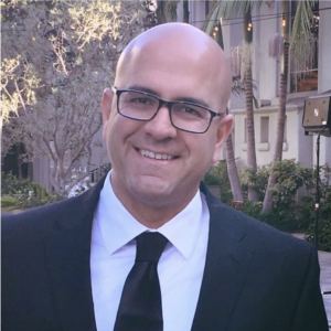 Lotfipour, Shahrdad, PhD
Mailing Address: Research objectives are to understand the mechanisms mediating addictive disorders. Special interest is on how gut, genes, brain, and behavior interact with environment (e.g., developmental drug exposure) to influence addiction. Focus is placed on gut bacteria, genetics, midbrain and limbic brain regions in modulating baseline and drug-induced brain and behavior associations. The goals of the research studies are to further our understanding of the impact of drugs of abuse on the developing and adult brain using animal models, with translational relevance to the human population. The information gathered aims to assist in the promotion of better prevention and intervention strategies for the reduction of addictive disorders in the future.
...view more
Research Areas: Drug Addiction, Neuropharmacology & Neurochemistry, Genetics & Epigenetics, Learning & Memory, Neurodegeneration & Neuropsychiatric Disorders, Neurodevelopment, Stem Cells & Plasticity/Regeneration, Systems/Circuit Neuroscience & Synaptic Transmission |
 Lur Gyorgy, PhD
Mailing Address: Neurons in the neocortex are bombarded with thousands of inputs representing different senses, memories of past events or internal goals. The Neuronal Signal Integration Lab at UC Irvine aims to understand how individual cells and networks of neurons integrate the information arriving from these different modalities. We combine opto- and pharmacogenetic strategies with electrophysiology and optical recording methods to manipulate and monitor neuronal activity. These tools allow us to ask what neuronal signals underpin complex behaviors like decision-making, and how adverse environments disrupt these information streams, leading to impaired cognitive function. for more information:
...view more
Research Areas: Learning & Memory, Systems/Circuit Neuroscience & Synaptic Transmission |
 Lyon, David C., PhD
Mailing Address: Organization and function of the visual system in healthy animal models and models of disease.
...view more
Research Areas: Systems/Circuit Neuroscience & Synaptic Transmission |
 Mahler, Stephen V., PhD
Mailing Address: In order to attain a natural reward like food, water, or sex, animals must know what and where rewards are, and how to get them. This is accomplished in part via the brain’s “reward circuitry,” aspects of which allow animals to recognize rewards when they are attained, learn about the circumstances in which they were attained, remember these circumstances when they are encountered in future, and generate appropriate motivated behavior at those times.
...view more
Research Areas: Drug Addiction, Neuropharmacology & Neurochemistry, Learning & Memory, Systems/Circuit Neuroscience & Synaptic Transmission |
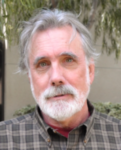 McNaughton, Bruce L., PhD
Mailing Address: I have made significant contributions to both basic neuroscience theory and research, and novel research technology (such as tetrode recording). Although my past theoretical and experimental work has focused largely on information coding and intrinsic dynamics of the hippocampus itself, my focus for the next decade will be on the question: what does the hippocampus do for the neocortex? How does hippocampal outflow integrate and shape cortical activity and representational structure? I am interested in the impact of hippocampal outflow on neocortical information processing, intrinsic dynamics, connectivity dynamics, and ‘semantic’ encoding. My guiding hypothesis is that hippocampus serves initially to link new information distributed in sparsely connected neocortical autoassociative modules and to orchestrate the offline creation of semantic knowledge from this episodic memory (see McClelland et al, 1995).To address questions related to this hypothesis I am using a range of neuroscience enabling technologies in rats and genetically modified mice (e.g., multi neuron ensemble recording, Ca++, voltage, and structural imaging).
...view more
Research Areas: Learning & Memory, Systems/Circuit Neuroscience & Synaptic Transmission |
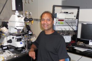 Metherate, Raju, PhD
Mailing Address: The long-term goal of my lab is to understand how nicotinic acetylcholine receptors (nAChRs) regulate neural processing in auditory cortex, and to develop nicotine-based drug therapies for auditory processing disorders. Our research accomplishments highlight novel cellular and systems-level mechanisms of nicotinic modulation. These mechanisms in auditory cortex and subcortical structures (notably the thalamocortical pathway) integrate to produce a remarkable result: systemic nicotine increases gain and sharpens receptive fields in primary auditory cortex (e.g., Askew et al. 2017). Going forward, the lab has two broad goals: first, to understand which nAChR subtypes and neuron sub-classes are responsible for the effects of nicotine at the systems and behavioral-cognitive levels. In particular, we wish to determine how the most striking of nicotine’s cellular actions—selective and robust excitation VIP interneurons (Askew et al. 2019)—contributes to nicotine’s physiological effects. A second broad goal is translational: to determine if nicotine’s effects may enable a first-ever drug treatment for central auditory processing disorders that involve diminished auditory attention. Given that systemic nicotine mimics the effects of auditory attention on receptive fields in A1 of humans and nonhuman primates, we wish to determine if nicotine may enhance auditory attention, in particular during aging and disorders associated with reduced auditory-cognitive function. To these ends our research focuses on mouse in vivo and in vitro electrophysiology and, in collaboration with other labs, nicotine’s effects in behaving animals and humans. Intskirveli, I. and Metherate, R. (2012) Nicotinic neuromodulation in auditory cortex requires MAPK activation in thalamocortical and intracortical circuits. Journal of Neurophysiology 107:2782-2793. PMCID: PMC3362282 Askew, C.E., Intskirveli, I. and Metherate, R. (2017) Systemic nicotine increases gain and narrows receptive fields in A1 via integrated cortical and subcortical actions. eNeuro 4:e0192-17.2017 1–18. PMCID: PMC5480142 Askew, C.E., Lopez, A.J., Wood, M.A and Metherate, R. (2019) Nicotine excites VIP interneurons to disinhibit pyramidal neurons in auditory cortex. Synapse 73:e22116. PMCID: PMC6767604 Pham C.Q., Kapolowicz M.R., Metherate R. and Zeng F.G. (2020) Nicotine enhances auditory processing in healthy and normal-hearing young adult nonsmokers. Psychopharmacology (Berl) 237:833-840. doi: 10.1007/s00213-019-05421-x. PMID: 31832719
...view more
Research Areas: Drug Addiction, Neuropharmacology & Neurochemistry, Learning & Memory, Neurodegeneration & Neuropsychiatric Disorders, Systems/Circuit Neuroscience & Synaptic Transmission |
 Monuki, Edwin S., PhD
Mailing Address: My research focus is the human choroid plexus. The choroid plexus is a secretory machine that makes the cerebrospinal fluid (CSF), forms the blood-CSF barrier, detoxifies harmful agents, and gates CNS immune cells. As such, the choroid plexus is a major and highly strategic interface between the body and brain with tremendous untapped potential for CNS studies and regenerative medicine applications. To tap into this potential, my lab leverages its patented technology for deriving choroid plexus epithelial cells from human pluripotent stem cells and its access to human choroid plexus samples from the UC Irvine autopsy service and Alzheimer’s Disease Research Center. Current approaches include developmental and disease modeling with isogenic iPSCs, single cell omics, and deep machine learning of 2D whole slide and 3D digital images. Other research activities include collaborative studies on forebrain development, brain malformations, neurodegenerations, and preclinical studies of human stem cell transplantation.
...view more
Research Areas: Human Neuroscience, Neurodegeneration & Neuropsychiatric Disorders, Neurodevelopment, Stem Cells & Plasticity/Regeneration |
 Ostlund, Sean, PhD
Mailing Address: Research Interests: Motivated behavior, Reward, Addiction and Substance Abuse, Dopamine, Decision Making
...view more
Research Areas: Drug Addiction, Neuropharmacology & Neurochemistry |
 Othy, Shivashankar, PhD
Mailing Address: Our goal is to uncover the principles of immune regulation in the central nervous system (CNS) at organismal, cellular, and molecular levels. We study the cellular and molecular mechanisms of regulatory (Treg) cells during autoimmune neuroinflammation using Multiple Sclerosis (MS)-like disease model in mice. Our research intersects immunology, cell biology, physiology, and neuroscience. We use a combination of advanced multiphoton imaging, fate-mapped reporters, biophysical tools, and functional genetics in animal models to study spatiotemporal dynamics of Regulatory Immunity in CNS. Our studies illuminate the basic mechanisms of how Treg cells resolve neuroinflammation (CNS autoimmunity) and promote neuronal repair pathways (Remyelination). We aim to develop of Treg-cell therapy to achieve site-specific remyelination of denuded axons (e.g., MS) and lay the foundation for the CNS-tissue repair potential of Treg cells for other neurodegenerative diseases such as Alzheimer’s disease. In the long term, we seek to broaden our understanding of how neuroinflammation is regulated; this will guide the development of new therapeutic strategies for neuroinflammatory and autoimmune diseases of the CNS.
...view more
Research Areas: Neuroimmunology, Neuroinflammation & Glia |
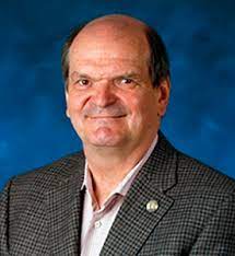 Palczewski, Krzysztof (Kris), PhD.
Mailing Address: Gene augmentation therapy for correcting mutations in the RPE65 gene is the first FDA-approved gene therapy for any genetically inherited disease. There is no doubt that further genetic editing represents a new frontier that will permit the rescue of a specific function disabled by a genetic mutation. However, the clinical translation of CRISPR-Cas9 technology has been impeded by its low editing efficiency by homology-directed repair (HDR) and substantial indel formation caused by double-stranded DNA (dsDNA) breaks. Base editing is a new and innovative alternative for correcting gene mutations. Cytosine and adenine base editors (CBEs and ABEs), an advanced CRISPR-Cas9-associated genome editing tool, enable conversion of a point mutation independent of Cas9-induced dsDNA breaks and HDR, thereby addressing the drawbacks of conventional CRISPR-Cas9 technology. Base editing is especially attractive as a method to correct mutations in key genes associated with phototransduction, the visual (retinoid) cycle and other cellular processes essential for maintaining
...view more
Research Areas: Genetics & Epigenetics, Neurodegeneration & Neuropsychiatric Disorders, Neurodevelopment, Stem Cells & Plasticity/Regeneration, Systems/Circuit Neuroscience & Synaptic Transmission |
 Pathak, Medha M., PhD
Mailing Address: The long-term goal of our lab is to understand how mechanical forces modulate neural stem cell fate in development and repair at molecular, cellular and organismal levels. We previously showed that the stretch-activated ion channel Piezo1 mediates mechanosensitive lineage specification of neural stem cells. Our studies revealed that Piezo1 activity in neural stem cells is modulated by matrix mechanics, and that it influences differentiation of the cells into neurons or astrocytes. Current work focuses on understanding (i) how Piezo1 detects and transduces matrix mechanical signals, (ii) how Piezo1 activity elicits gene expression changes, (iii) how Piezo1 may shape neural development, and (iv) how Piezo1 may be involved in certain disease states. We use a multi-disciplinary approach, combining ideas and techniques from ion channel biophysics, cell biology, optical imaging, stem cell biology and bioengineering.
...view more
Research Areas: Neurodevelopment, Stem Cells & Plasticity/Regeneration |
 Piomelli, Daniele, PhD
Mailing Address: My lab studies the neurobiology and pharmacology of the endocannabinoids and other bioactive lipids. Our approach combines molecular and cellular biology, system biology and medicinal chemistry to discover transformative chemical probes that target lipid-signaling pathways. We were the first to elucidate pathways involved in the formation and deactivation of brain endocannabinoids, uncover important physiological functions of these compounds, and identify pharmacological agents that interfere with their deactivation. Agents discovered by my group have been broadly used by the scientific community and have inspired preclinical and clinical research in many labs of both academe and industry.
...view more
Research Areas: Drug Addiction, Neuropharmacology & Neurochemistry, Neurodegeneration & Neuropsychiatric Disorders |
 Rose, Matthew, MD, PhD.
Mailing Address: We study the gene networks driving neuronal diversity during brainstem development to uncover why only specific subsets are differentially affected in a given human neurologic disease. The brainstem controls multiple critical motor and sensory functions, including eye movements, facial expression, speech, hearing, proprioception, arousal, and breathing. Disruption of these functions can lead to profound deficits including in childhood social interaction. The anatomy and gene circuits in the brainstem are highly conserved in mice, providing an ideal model system to investigate neuronal specification and axon growth and guidance in both health and disease. We combine neurodevelopmental tools, 3D imaging, multi-omics/bioinformatic approaches, molecular/synthetic biology, and mouse models of human disease to define developmental differences among various neuronal populations. Through a single cell and spatial transcriptomic atlas of developing motor neurons, we have identified unique genetic fingerprints of each motor neuron population, including novel markers of spatially and temporally distinct subpopulations that contribute to specific aberrant nerve branches in mouse models of childhood disorders. This work provides a new toolbox to study differential neuronal vulnerability in disease.
...view more
Research Areas: Genetics & Epigenetics, Human Neuroscience, Neurodegeneration & Neuropsychiatric Disorders, Neurodevelopment, Stem Cells & Plasticity/Regeneration, Systems/Circuit Neuroscience & Synaptic Transmission |
 Sajjadi, S. Ahmad, PhD
Mailing Address: In my lab, we study degenerative brain diseases using a combination of neuro-imaging analysis, neuropsychological assessments, and neuro-pathology. We have a particular emphasis on degenerative brain diseases that come into differential diagnosis of Alzheimer’s disease. We especially focus on the diagnosis of their underlying proteinopathies.
...view more
Research Areas: Human Neuroscience, Neurodegeneration & Neuropsychiatric Disorders |
 Schechtman, Eitan, PhD
Mailing Address: The Cognitive Neuroscience of Sleep lab (or CogNoS for short) uses neuroimaging, behavioral manipulations, and computational methods to explore memory reactivation during sleep, which broadly impacts cognition, emotion, and health. Exploring the mechanisms through which sleep shapes human behavior requires a multi-disciplinary approach, combining neuroscientific, psychological, and computational methods. Our lab combines novel techniques to selectively bias memory reactivation; machine-learning algorithms to decipher memory-related content from neural data; and neuroscientific methods for monitoring brain connectivity and rhythms in different regions and timescales. Using this state-of-the-art methodological framework, the lab hopes to reveal the neural infrastructure through which sleep transforms memories, and how these dynamics may be harnessed for improving wellbeing in healthy and clinical populations
...view more
Research Areas: Human Neuroscience, Learning & Memory |
 Schilling, Thomas F., PhD
Mailing Address: Our laboratory uses genetics and molecular biology to study pattern formation in the zebrafish embryo. The rapid development and simple anatomy of this teleost embryo, together with recently developed techniques for reverse genetics and a complete genome sequence, make zebrafish a powerful molecular genetic system for studying the mechanisms of development. We are interested in how gene functions translate into cell behaviors and the formation of tissues and organs. We focus on two main areas relevant to the nervous system: 1) cell interactions and formation of segments along the anterior-posterior axis of the hindbrain in the central nervous system, 2) migration of neural crest cells and how they form the neurons and glia of the peripheral nervous system. We combine experimental and computational/systems biology approaches to these two aspects of neural development. Our most recent studies use genome-wide gene expression profiling, such as single cell RNA-sequencing, to address how cells in the hindbrain and PNS obtain their fates.
...view more
Research Areas: Genetics & Epigenetics, Neurodevelopment, Stem Cells & Plasticity/Regeneration |
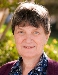 Seiler, Magdalene J. Ph.D.
Mailing Address: Retinal progenitor sheet transplantation and demonstration of vision improvement in models of retinal degeneration and ultimately in retinal degeneration patients (Retinitis Pigmentosa, age-related macular degeneration)
...view more
Research Areas: Neurodegeneration & Neuropsychiatric Disorders, Neurodevelopment, Stem Cells & Plasticity/Regeneration |
 Stark, Craig, PhD
Mailing Address: Dr. Stark’s research investigates the neural bases of human long-term memory. He uses multiple forms of MRI imaging (functional, structural, diffusion, and spectroscopy) in conjunction with experimental psychology and studies of aging and dementia to investigate how the neural systems supporting these various types of memory operate and interact. Particular emphasis is placed on understanding the human hippocampus and other components of the medial temporal lobe.
...view more
Research Areas: Human Neuroscience, Learning & Memory, Neurodegeneration & Neuropsychiatric Disorders |
 Steward, Os, PhD
Mailing Address: The overall focus of my research is on how nerve cells create and maintain their connections with each other, and how synapses are modified by experience and after injury (synaptic plasticity). One component of the research follows on our discovery of the fundamental mechanism underlying local protein synthesis at synapses, which revealed a previously unknown aspect of neuronal cell biology. We were the first to advance the now well-accepted idea that local protein synthesis at synapses plays a critical role in enduring forms of synaptic plasticity and memory. Subsequent studies contributed to the understanding of mechanisms underlying the transport of mRNA into dendrites, selective docking of mRNA at active synapses, and activity-dependent mechanisms of mRNA decay. This core idea drove research in the fields of neuroscience and cell biology that now involves hundreds of other labs, leading to our current understanding that local protein synthesis plays a pivotal role in normal synaptic function, and that disorders of synaptic protein synthesis may underlie important developmental disorders including Fragile-X Mental Retardation Syndrome and autism spectrum disorders. A major component of my research over the past 2 decades has been on axon regeneration following spinal cord injury (SCI). Our research team collaborated with Zhigang He’s team and others on a breakthrough report showing that conditional deletion of PTEN enabled unprecedented regeneration of corticospinal tract (CST) axons after SCI. Since then, we have developed approaches to enable regeneration via AAVshRNA-mediated knockdown and demonstrated that CST regeneration is accompanied by recovery of skilled motor function. A major recent research focus has been on the design and deployment of AAV vectors to deliver gene modifying cargoes to enhance regenerative capacity after injury. Of particular interest are AAVs that undergo retrograde transport (retro-AAVs), which can deliver cargoes to neurons in the brain that give rise to spinal pathways. Our laboratory uses a variety of techniques ranging from molecular biology, especially in situ hybridization, immunohistochemistry, electron microscopy, light microscopy including light sheet microscopy, neurophysiology, and behavioral assays of sensory and motor function. Our research is supported by grants from the National Institutes of Health and private donations. Training of graduate students and postdoctoral fellows is very important to me, and I’m greatly honored to have received the NINDS Story Landis Award for Outstanding Mentorship in 2020.
...view more
Research Areas: Learning & Memory, Neurodevelopment, Stem Cells & Plasticity/Regeneration, Systems/Circuit Neuroscience & Synaptic Transmission |
 Swarup, Vivek, PhD
Mailing Address: Our lab uses multi-omic approaches to understand molecular mechanism of neurodegenerative diseases like Alzheimer’s disease and Fronto-temporal dementia (FTD). We use functional genomics approaches to better understand the role of distinct cell-types in disease pathophysiology to improve the design of therapeutic interventions for AD and FTD. Current transcriptomic approaches can powerfully investigate quantitative molecular phenotypes and pathways underlying disease progression in a genome-wide manner. Yet, they lack the specificity needed to comprehend the role of cell-type specific changes in disease pathophysiology. Our work will identify the molecular mechanisms underlying neurodegeneration and investigate how they differ from normal aging. Using single-cell multi-omic approaches coupled with bulk tissue sequencing, we hope to directly answer some of these questions.
...view more
Research Areas: Genetics & Epigenetics, Neurodegeneration & Neuropsychiatric Disorders, Neuroimmunology, Neuroinflammation & Glia |
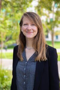 Thompson-Peer, Katherine L., PhD
Mailing Address: Investigating how neurons respond and recover after injury The Thompson-Peer lab explores how neurons recover from injury in vivo, and how this process is similar to and different from normal development. At the most fundamental level, a neuron receives information along dendrites, and sends information down an axon to synaptic contacts. Dendrites can be injured by traumatic brain injury, stroke, and many forms of neurodegeneration, yet while the factors that control axon regeneration after injury have been extensively studied, we know almost nothing about dendrite regeneration. Our long term research goal is to understand the cellular mechanisms of dendrite regeneration after injury. Our previous work found that the sensory neurons in the fruit fly Drosophila peripheral nervous system exhibit robust regeneration of dendrites after injury and used this system to explore central features of dendrite regeneration in developing animals, young adults, and aging adults. We have observed that after injury, neurons regrow dendrites that recreate some features of uninjured dendrites, but are unable to reconstruct an entire arbor that perfectly mimics an uninjured neuron. Moreover, there are mechanistic differences between the outgrowth of uninjured neurons versus the regeneration of dendrites after injury: dendrite regeneration is uniquely dependent on neuronal activity, ignores cues that constrain and pattern normal dendrite outgrowth, and confronts a mature tissue environment that is different from what a developing neuron would encounter. These challenges are significantly exacerbated when neurons in aging animals attempt to recover from injury. Current and future projects will deepen our knowledge about dendrite regeneration to create a new framework for understanding how neurons recover from injury in both young and aging animals.
...view more
Research Areas: Genetics & Epigenetics, Neurodevelopment, Stem Cells & Plasticity/Regeneration |
 Thompson, Leslie M., PhD
Mailing Address: Huntington’s disease (HD) is a devastating, inherited neurological disorder that causes psychiatric, cognitive and movement deficits.The Thompson laboratory is actively engaged in investigating the fundamental molecular and cellular events that underlie how the mutant HD gene causes degeneration of specific brain cell populations to induce motor and cognitive decline and premature death of patients with the ultimate goal to develop new therapeutic approaches. Several potential drug targets that have emerged are in various phases of target validation and preclinical and clinical trials, including stem cell-based therapies. The laboratory also focuses on understanding casual mechanisms that underlie HD and Amyotrophic Lateral Sclerosis and more recently X-linked Dystonia-Parkinsonism with the goal of developing treatments for the disease. The research benefits from the integrated use of patient iPSCs and mouse models of disease together with the studies of RNA biology and network-based bioinformatics.
...view more
Research Areas: Genetics & Epigenetics, Neurodegeneration & Neuropsychiatric Disorders, Neurodevelopment, Stem Cells & Plasticity/Regeneration, Neuroimmunology, Neuroinflammation & Glia |
 Tombola, Francesco, PhD
Mailing Address: My lab studies the molecular sensors that allow cells to detect and respond to physical cues in their environment, including electrical and mechanical stimuli. Cells use these sensors to adapt to and modify their microenvironment, and to communicate with each other. In the nervous system, we are particularly interested in how resident immune cells communicate with neurons and glia. We focus on ion channels that are activated by changes in membrane potential and are permeable to protons or potassium, and on channels that are activated by increases in membrane tension and are permeable to calcium. We utilize a variety of techniques, comprising electrophysiology, imaging, biochemistry and molecular biology methods, kinetic modeling, and molecular dynamics simulations, to study: 1) how these channel proteins work and are modulated by intra- and extracellular signals, and by pharmacological treatments, and 2) how their activity affects cellular physiology and pathophysiology. Some of the functions of the sensors we study are known. For example, the Hv1 channel regulates the production of reactive oxygen species in microglial cells and has proinflammatory activity in the context of ischemic stroke, whereas the Piezo1 channel helps neural stem progenitor cells make fate choices. Other functions, on the other and hand, are completely unknown and await enthusiastic and ambitious graduate students to be discovered.
...view more
Research Areas: Neurodevelopment, Stem Cells & Plasticity/Regeneration, Neuroimmunology, Neuroinflammation & Glia |
 Villalta, Sergio ‘Armando’, PhD
Mailing Address: The Villalta laboratory is directed towards understanding how immune cells contribute to tissue injury and repair in degenerative and autoimmune diseases.
...view more
Research Areas: Neurodegeneration & Neuropsychiatric Disorders |
 Walsh, Craig M., PhD
Mailing Address: Our research is focused on the impact of adaptive immunity, primarily T cells, on the CNS. We have two major projects in this area. First, we are studying the effect of an immune cell population called regulatory T cells (Tregs) on remyelination and CNS regeneration in models of demyelinating diseases related to multiple sclerosis. In this work, we have found that neural stem cells provoke the entry of Tregs into the CNS, helping to dampen inflammation and promote remyelination and are studying how such cells might be used to treat multiple sclerosis. Second, we are interested in the role(s) of adaptive immune cells (T and B cells) in Alzheimer’s Disease (AD), and are exploring the effect of a loss of adaptive immunity on AD progression in mouse models. Our work is geared toward understanding the crosstalk between the CNS and immune system.
...view more
Research Areas: Neuroimmunology, Neuroinflammation & Glia |
 Watanabe, Momoko, PhD
Mailing Address: Our project focuses on the development of highly efficient and reproducible brain organoid methods to investigate the origins of neural circuits and model neurodevelopmental disorders. We have contributed to set up cortical-subcortical fused-organoids to identify neural oscillations and establish a platform to examine interneuronopathy for neurodevelopmental disorders. Recently, we have worked on a project to determine the transcriptomic signatures of human pluripotent stem cells that predict efficient and reproducible cortical organoid formation. We have set out in our near future work to address three main aims: 1) create a powerful organ-on-a-chip systems to improve the established forebrain organoids, 2) develop brain organoid models for neurodevelopmental disorders, and 3) define the microcircuit properties of cortical organoids as a model system for human brain activities.
...view more
Research Areas: Human Neuroscience, Neurodegeneration & Neuropsychiatric Disorders, Neurodevelopment, Stem Cells & Plasticity/Regeneration |
 Weiss, John H., PhD
Mailing Address: John H. Weiss, MD, Ph.D., Professor
...view more
Research Areas: Neurodegeneration & Neuropsychiatric Disorders |
 Wood, Marcelo, PhD
Mailing Address: Long-term memory storage is an essential process to human life. Without long-term memory, we would not be able to remember our pasts, interpret our present, or predict our future. We would have little personal identity, and functioning in a world that continues to grow in complexity would be impossible. Our research goal in the Wood lab is to understand the molecular mechanisms underlying long-term memory processes and drug-seeking behavior. It has long been known that transcription is required for a learning event to be encoded into long-term memory. Successful transcription of specific genes required for long-term memory processes involves the orchestrated effort of not only transcription factors, but also very specific enzymatic protein complexes that modify chromatin structure. Chromatin modification (Barrett and Wood 2008) and remodeling (Vogel-Ciernia and Wood 2014) are two major epigenetic mechanisms that are essential for certain long-term forms of synaptic plasticity and memory. The best-studied form of chromatin modification in the learning and memory field is histone acetylation, which is regulated by histone acetyltransferases (HATs) and histone deacetylases (HDACs). Our lab primarily works on the HAT called CBP (e.g. Barrett et al., 2011), which we found to be essential for long-term memory formation, and HDAC3, which we have demonstrated to be a critical negative regulator of long-term memory formation (e.g. McQuown et al., 2011; Kwapis et al., 2018) and drug-seeking behavior (e.g. Rogge et al., 2011; Malvaez et al., 2013). One of the alluring aspects of examining chromatin modification and remodeling in modulating transcription required for long-term memory processes is that these modifications may provide transient and potentially stable epigenetic changes in the service of activating and/or maintaining transcriptional processes, which in turn may ultimately participate in the molecular mechanisms required for neuronal changes subserving long-lasting changes in behavior. As an epigenetic mechanism of transcription, chromatin modification and remodeling have been shown to maintain cellular memory (e.g. cell fate) and may underlie the strengthening and maintenance of synaptic connections required for long-term changes in behavior (Campbell and Wood, 2019). Indeed, we have demonstrated that inhibition of HDACs can modulate memory processes in fascinating ways (e.g. Stefanko et al., 2009; Malvaez et al., 2013; Lattal and Wood 2013). Current projects in the lab focus on several epigenetic mechanisms (e.g. histone modification, nucleosome remodeling, DNA methylation) and their role in regulating dynamic and coordinated gene expression profiles required for long-lasting forms of synaptic plasticity, memory, and drug-seeking behavior in the young and aged brain. We use molecular, genetic, viral, pharmacological and behavioral approaches in genetically modified animals and tissue culture. Research in the lab is supported by the National Institute on Drug Abuse, the National Institute on Aging, the National Institute of Mental Health, as well as private foundations and industry partners.
...view more
Research Areas: Drug Addiction, Neuropharmacology & Neurochemistry, Genetics & Epigenetics, Learning & Memory, Neurodegeneration & Neuropsychiatric Disorders, Systems/Circuit Neuroscience & Synaptic Transmission |
 Xu, Xiangmin, PhD
Mailing Address: Our research program focuses on neural circuit mechanisms that underlie sensory perception, learning and memory, and cognition. Using mouse models, we have made significant advances by showing cell-type specific and molecular signaling mechanisms underlying critical period synaptic plasticity. We demonstrated that the critical period window can be re-opened in adults to restore plasticity using targeted molecular manipulation, which has a potential for guiding new amblyopia treatments. We have extended our studies to other brain regions including prefrontal cortex and hippocampus towards our goal to determine whether neural plasticity can be manipulated for neural circuit functional repairs in brain disorders. Our newest direction is to apply our findings towards treating Alzheimer’s disease by pursuing circuit manipulation-based strategies in AD mouse models.
...view more
Research Areas: Learning & Memory, Neurodevelopment, Stem Cells & Plasticity/Regeneration, Systems/Circuit Neuroscience & Synaptic Transmission |
 Yassa Michael A, PhD
Mailing Address: My laboratory is interested in how the brain learns and remembers information, and how learning and memory mechanisms are altered in aging and neuropsychiatric disease. The central questions in our research are: What are the neural mechanisms that support learning and memory? We combine these approaches with more traditional psychophysics including measurements of galvanic skin response (skin conductance), heart rate variability, and eye tracking. We are also working with collaborators to develop novel platforms for cellular resolution functional imaging in awake, behaving animals using novel MRI tracers. Finally, we are actively developing and testing several pharmacological and nonpharmacological cognitive enhancement interventions in older adults at risk for dementia, including studies of physical exercise.
...view more
Research Areas: Human Neuroscience, Learning & Memory, Neurodegeneration & Neuropsychiatric Disorders, Systems/Circuit Neuroscience & Synaptic Transmission |
 Zeng, Fan-Gang, PhD
Mailing Address: Founded by Fan-Gang Zeng in 2000, the Hearing and Speech (HESP) laboratory at UC Irvine has been conducting basic and translational research in the following areas. (1) Understand mechanisms underlying normal and pathological hearing, (2) Understand relationships between hearing loss and cognitive decline, and (3) Find safe and effective treatment (e.g., electric stimulation, nicotine) for hearing loss, tinnitus and hyperacusis. The HESP lab is part of the UC Irvine Center for Hearing Research (hearing.uci.edu) and has provided research opportunities for a diverse group from high school and community college students to medical residents and visiting scholars. The HESP lab participates in formal doctoral and post-doctoral training programs in Anatomy and Neurobiology, Biomedical Engineering, Cognitive Sciences and Otolaryngology – Head and Neck Surgery. HESP lab members have also been actively involved in community service and outreach programs such as Lions Clubs International, American Tinnitus Association, the California State Summer School for Math & Science (COSMOS), and the UC Irvine Summer PreMed Program.
...view more
Research Areas: Human Neuroscience, Systems/Circuit Neuroscience & Synaptic Transmission |
